Engaged in manufacturing and exporting Botany Anatomy Models, Scientific Botany Models, Educational Botany Models, Laboratory Botany Models.
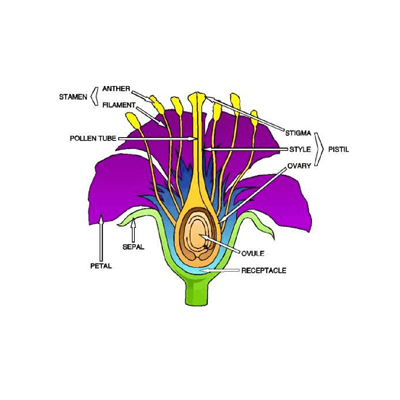
Model of typical flower, all parts detachable, ovary with a single ovule inside, mounted on base with key card.
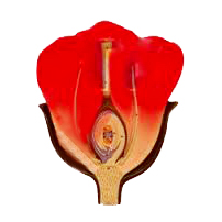
Model of L.S. flower, mounted on base with key card.
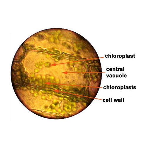
Enlarged many times, showing the microscopic structures, mounted on base with key card.
An electron microscopic structure, showing a portion of wall, ectoplast, endoplast, tonoplast, vacuoles nuclear structure, plastids, mitochondria etc, mounted on base with key card.
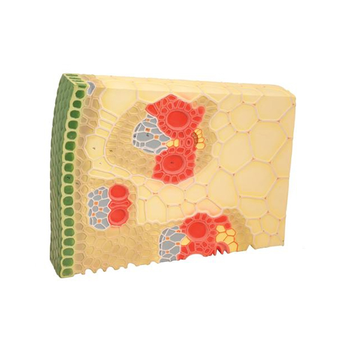
Model shows the transverse and longitudinal section of a dicotyledonous stem in which case the cambium ring has been formed but no secondary growth has yet taken place. Showing epidermis, lentice, cork layer, cork cambium, cortical parenchyma, starch sheath medullary rays, phloem, sieve plate, sieve tube, phloem parenchyma and inter fasicular cambium, Xylem, pitted vessels pith etc, mounted on bas.
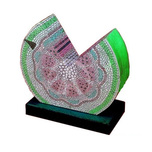
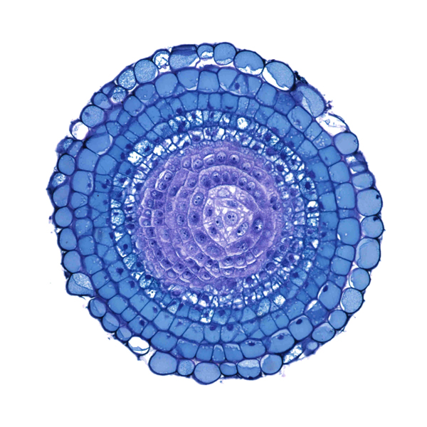
Showing various tissues vascular bundles in transverse section of a dicot stem of sunflower, mounted on base with key card.

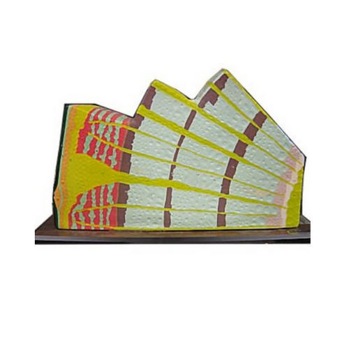
Exhibits the various tissues and scattered closed and collateral vascular bundles in transverse and longitudinal sections in maize. The large pitted vessels, spiral and annular vessels show the anatomy of monocot stem, mounted on base with key card.
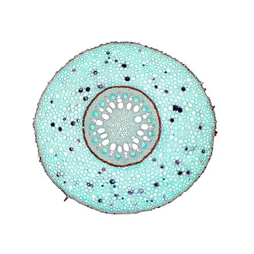
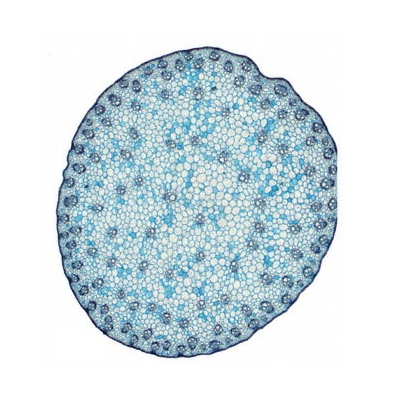
Showing various tissues vascular bundles in transverse section of monocot stem of maize, mounted on base with key card.
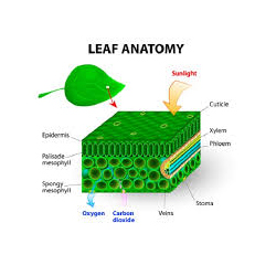
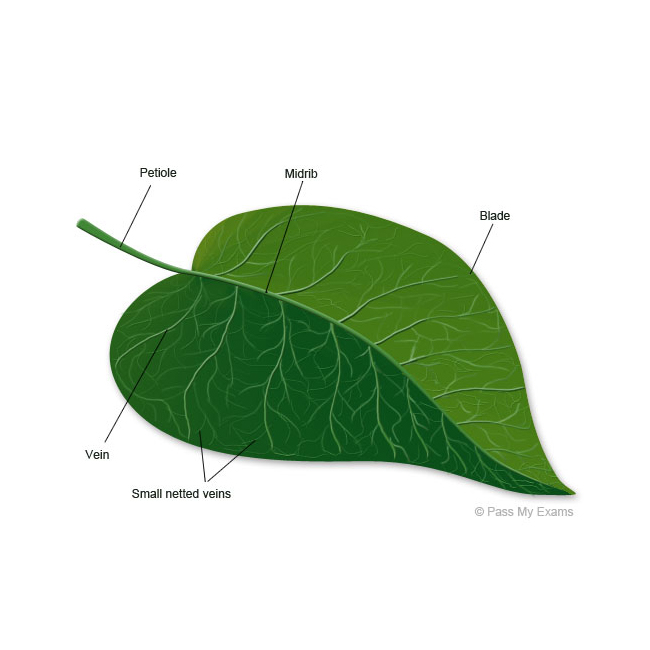
3-Dimensional model of leaf showing detailed structure of transverse section and longitudinal section, mounted on base with key card.

Showing details of typical mesophytic leaf, mounted on base with key card.
Showing details of internal structure of typical monocot leaf, mounted on base with key card.
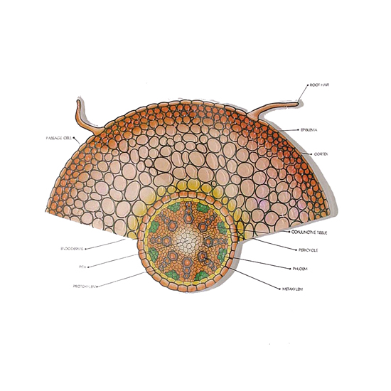
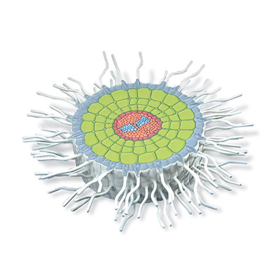
Showing complete internal details of root, mounted on base with key card.
Model250 times full sizes, Bean (Phaseolus vulgaris), mounted on base with key card.
Model shows the pointed growth resulting from an apical cell with the cells which branch out spirally from point of vegetation. Root shown in longitudes & transverse section, detachable crown of the root removable, mounted on base with key card.
OPEN COLLATERAL CONDUCTING BUNDLE OF DICOTYLE: Plant enlarged approx. 550 times, mounted on base with key card.
Polygonum type, enlarged 300 times, mounted on base with key card.
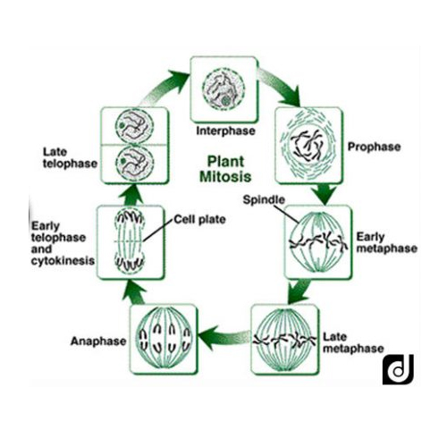
Set of 10 models showing all the stages of karyokinesis and cytokinesis form metabolic cell of the formation, mounted on base with key card.
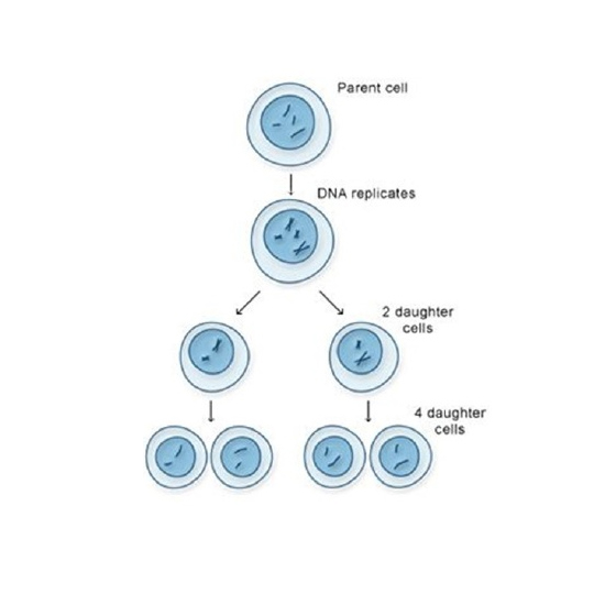
Set of 16, an entirely new designed model according to recent concept of chromosome changes from the resting nucleus to the formation of 4 daughter cells complete set, mounted on base with key card.
.jpg)
Model showing the germination of monocot plant (maize) seeds of development, mounted on base with key card.
.jpg)
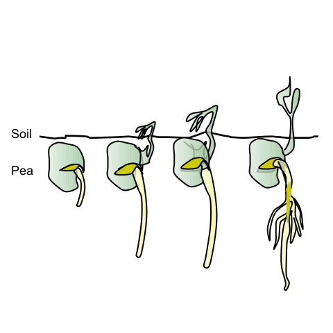
Model showing germination of dicot plant (pea) seeds of development, mounted on base with key card.
Model showing the detailed structure of leaf, mounted on base with key card.
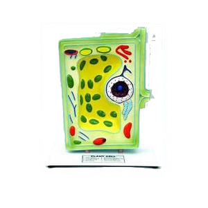
Model of a typical plant cell, magnified 50 million times. Shows the detailed structure including ectoplasm, cytoplasm, endoplasmic recticulum, vacuoles etc. Nucleus is removable, mounted on base with key card.

Model 40 times enlarged, showing the cross section of maize pant�s monocotyledonous stem (zea mays), mounted on base with key card.
Model of white mustard (sinapis alba) showing the absorption zone of a dicotledonous plant, mounted on base with key card.
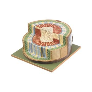
Model 40 times enlarged, showing the cross and longitudinal section through a 3-year old branch of lime tree (tilia platyphyllos), mounted on base with key card.
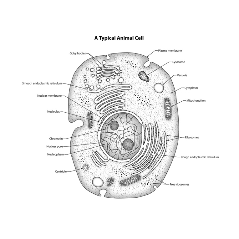
Model showing the ultra-structure of a cell. Set of 3 models, mounted on base with key card.
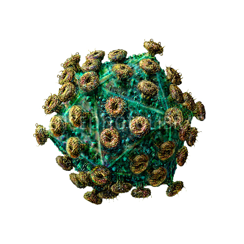
Model of HIV virus is enlarged millions of times shows the outer lipid membrane with protein structures, and the internal nucleus which contains the viral hereditary matter (RNA). The nucleus is removable and condoms can be put underneath to provide a message regarding measures to take in protecting against HIV infections, mounted on base with key card.
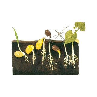
Eight-piece model for comparison of three different types of germination methods: monocot, dicot and gymnosperm.
Ambala Science Market © all rights reserved